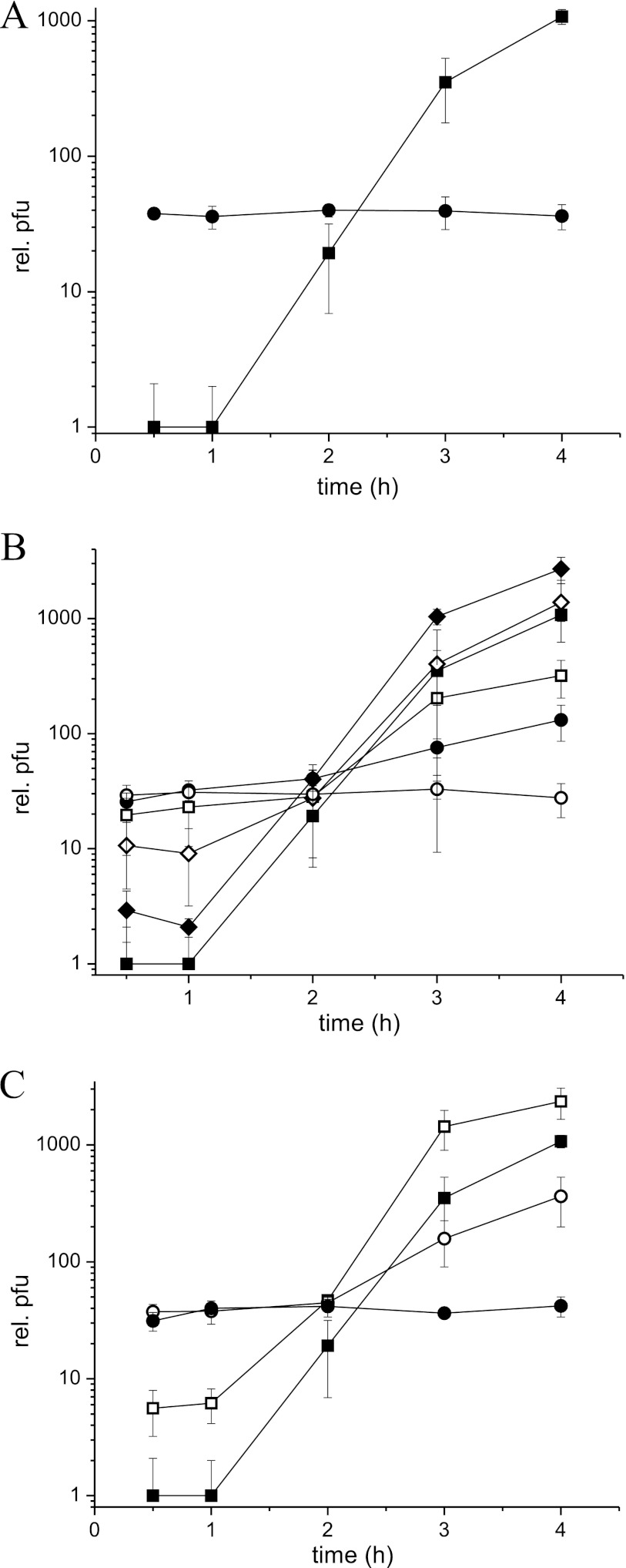
Fig 3.
Bacteriophage growth after addition of phage 7-7-1 to bacterial cultures. (A) Agrobacterium sp. H13-3 (■) and Agrobacterium tumefaciens (●). (B) Agrobacterium sp. H13-3 wild-type cells (■) and flagellar mutant cells: ΔflaA (□), ΔflaB (◆), ΔflaD (♢), ΔflaBD (●), and ΔflaABD (○). (C) Agrobacterium sp. H13-3 wild-type cells (■) and motility mutant cells; motAE98K (○), motAE98Q (□), and ΔmotA (●). Cultures were adjusted to an OD600 of 0.3, corresponding to 3.5 × 10⁸ cells/ml, and phage 7-7-1 was added at an MOI of 0.01. After 15 min of phage adsorption, the mixtures were diluted 1:1,000 and incubated further at 30°C. Samples were taken at 1-h intervals, and PFU were determined by the soft agar layer technique. Each data point represents the average from three independent experiments, and error bars represent standard deviations.

