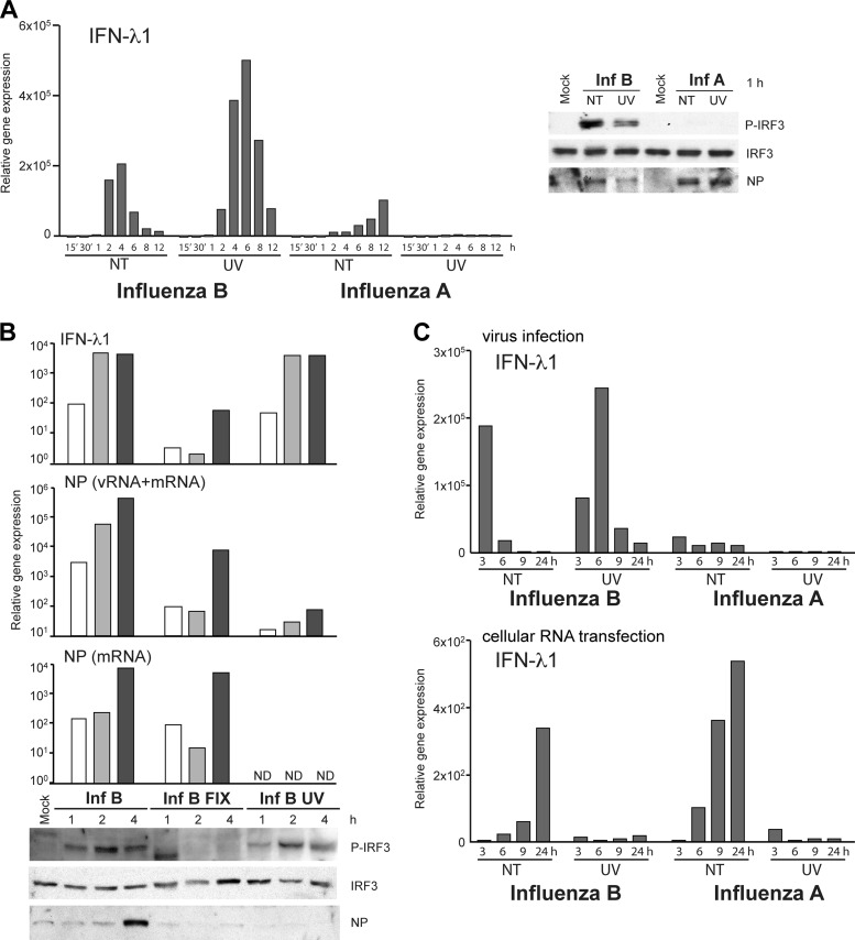
Fig 8.
Viral transcription is dispensable for influenza B virus-triggered IFN induction. (A) UV-irradiated or nontreated (NT) infectious influenza A/89 and B/97 viruses were used to infect moDCs at an MOI of 5. Infected cells were collected at different times, total cellular RNA was isolated, and IFN-λ1 gene expression was measured by qPCR. Data are representative of four independent experiments. For the analysis of IRF3 phosphorylation, cells were infected with high doses of UV-treated or live viruses (MOI of 30). Cells were collected, and cellular proteins were separated on 10% SDS-PAGE gels and immunoblotted with anti-P-IRF3, anti-IRF3, and influenza A and B virus NP-specific antibodies. (B) Influenza B/97 viruses were fixed with 3% paraformaldehyde and irradiated by UV light or were left untreated prior to the infection of moDCs with a dilution that corresponds to an MOI of 5. The gene expression levels of IFN-λ1 and viral NP vRNA or mRNA were measured from total cellular RNA and are shown as fold increases over the value for the mock sample. Phosphorylated and total IRF3 and viral NP levels in whole-cell lysates were visualized by immunoblotting. ND, not detected. (C) Cellular RNA from a virus infection experiment similar to that described above for panel A was analyzed for IFN-λ1 gene expression by qPCR (top) and further transfected into a fresh set of moDCs, and RNA-induced IFN-λ1 gene expression at 4 h posttransfection was analyzed by qPCR (bottom).

