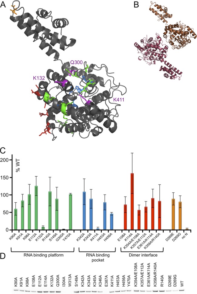
Fig 3.
In vivo effects of site-directed CCHFV N protein mutants. (A) Residues selected for mutation and testing in the minireplicon system are highlighted on the structure. Residues in the RNA binding platform are highlighted in green, residues that comprise the RNA binding pocket are shown in blue, the possible dimer interface is shown in red, and D269 of the DEVD motif is shown in orange. Alterations of residues K132, Q300, and K411 abrogated minigenome activity, and these residues are shown in magenta. (B) Ribbon representation of two protomers within the crystal lattice, suggesting the putative dimer interface investigated using the minireplicon system. (C) Histogram showing reporter gene expression for mutants, colored as described above, normalized to that of wild-type (WT) N. (D) Western blot analysis to assess relative expression levels of N protein mutants from equivalent quantities of cells expressing the minireplicon system, using a polyclonal antibody raised against the CCHFV N protein.

