
Fig 5.
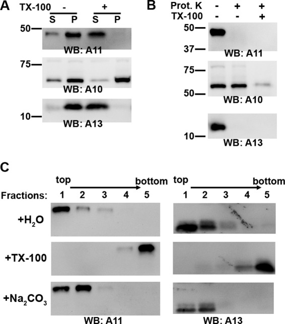
A11 associates with membranes during normal VACV replication. HeLa cells were infected with WT VACV at an MOI of 5 PFU/cell for 8 h, and the cell lysates were fractionated by ultracentrifugation on a 5 to 25% continuous iodixanol gradient as described for Fig. 3. The main fractions containing A11 (fractions 10 to 12) were pooled together and treated as follows. (A) The A11 fraction was mixed with Triton X-100 at a final concentration of 1% (+) or with water (-), layered on top of a 0.5 M sucrose cushion, and centrifuged at 100,000 × g for 2 h. The pellet (P) and TCA-precipitated supernatant (S) were analyzed by Western blotting. The positions and masses (in kilodaltons) of marker proteins are indicated on the left. (B) The A11 fraction was subjected to limited proteinase K digestion in the presence or absence of 1% Triton X-100 and analyzed by Western blotting. (C) The A11 fraction was incubated for 30 min with water (H2O), with Triton X-100 (TX-100) at a final concentration of 1%, or with Na2CO3 (pH 11.5) at a final concentration of 100 mM. The iodixanol concentration of the sample was adjusted to 30%. One milliliter of the sample was placed at the bottom of a centrifuge tube and overlaid with 3 ml of 25% iodixanol and 0.5 ml of 5% iodixanol. Ultracentrifugation was performed at 200,000 × g for 2 h. Fractions of the gradient were precipitated with TCA and analyzed by Western blotting. The direction of the gradient is indicated above the lanes.