
Fig 5.
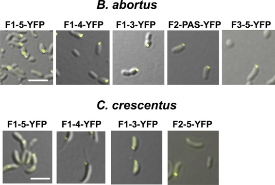
Localization assays. Shown are fragments of PdhS-YFP fusions that are able to polarly localize in B. abortus (upper panels) and in C. crescentus (lower panels). All fluorescence micrographs are merged differential interference contrast (DIC) and YFP images. Scale bar, 3 μm.