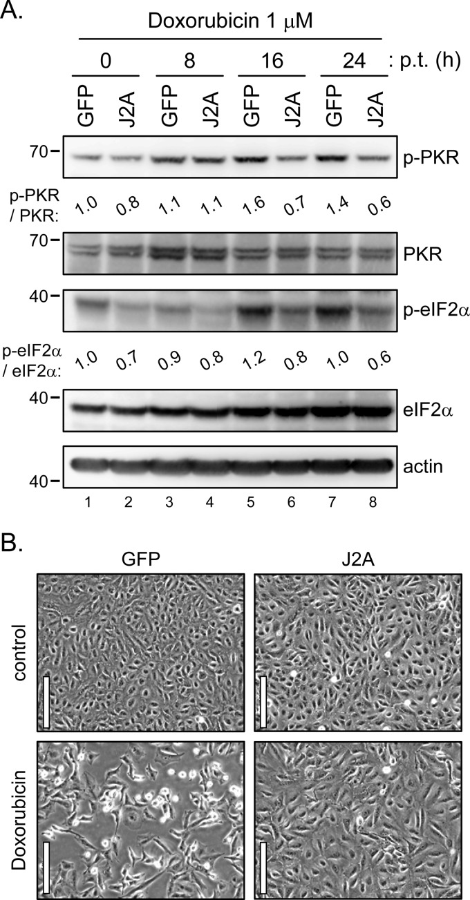
Fig 6.
JNS2A reduced the doxorubicin-triggered PKR activation and cell death. (A) Stable JNS2A and GFP control cells treated with doxorubicin (1 μM) for various times were harvested for Western blot analysis with the indicated antibodies. The bands intensities were quantified by MetaMorph, and the relative levels for the indicated proteins are shown. (B) Cell morphology of GFP/A549 and J2A/A549 cells after doxorubicin (1 μM) treatment for 32 h by phase-contrast microscopy. Scale bar, 100 μm.

