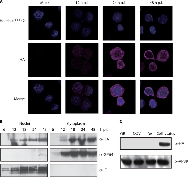
Fig 8.
Localization of Ac34 in vHA:Ac34-infected cells and purified virions. (A) Immunofluorescence analysis of the subcellular localization of Ac34. Sf9 cells were infected with vAcWT (Mock) or vHA:Ac34 at an MOI of 5 TCID50/cell. At various time points, the cells were fixed, probed with mouse monoclonal anti-HA antibody to detect HA:Ac34, and visualized using Alexa 555-conjugated goat anti-mouse antibody (violet). Nuclei were stained with Hochest33342 (blue). All of the images are shown at the same magnification. (B) Western blotting of the subcellular localization of Ac34. Sf9 cells were infected with vHA:Ac34 at an MOI of 5 TCID50/cell, and cytoplasmic and nuclear fractions were prepared at the indicated time points. The samples were subjected to immunoblotting with anti-HA, anti-IE1, or anti-GP64 antibodies. (C) Analysis of Ac34 in purified virions. Lysates of vHA:Ac34-infected cells and purified vHA:Ac34 virions (BVs, ODVs, and OBs) were subjected to immunoblotting using anti-HA and anti-VP39 antibodies.

