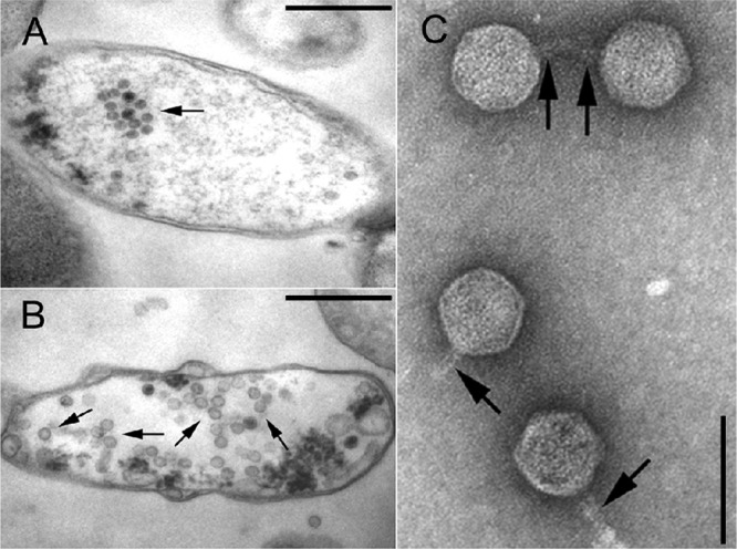
Fig 4.

TEM of P13374 induction from lysogenic strain TPE2364. Ultrathin sections of two bacterial cells (TPE2364) with maturating virion particles within the cytoplasm indicated by arrows (bars, 500 nm). The cells show signs of bacteriolysis (loss of cytoplasm) (A) and damaged cell walls, as well as the formation of vesicles or opaque clumps (B). (C) TEM of CsCl-purified, negatively stained P13374 particles released by strain TPE2364 (bar, 100 nm). Four phages with a short tail (arrows) and a hexagonal head are shown. In the upper part, two phages with tail-to-tail interactions are shown.
