Fig 1.
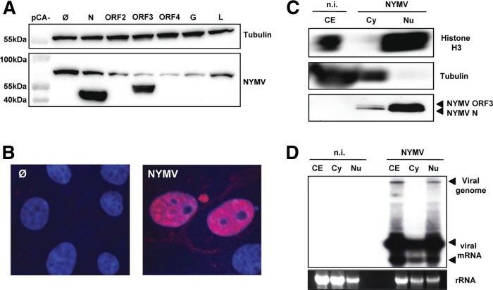
NYMV replicates in the nucleus. (A) Specificity for NYMV antigens of serum from NYMV-infected mice. 293T cells were transfected with expression vectors encoding the indicated NYMV proteins or the empty vector as a control (Ø). At 24 h posttransfection the cells were lysed, and Western blot analysis was performed. Antibodies against β-tubulin were used to control for equal loading of the gel. (B) Immunofluorescence analysis. Infected (NYMV) and uninfected (Ø) Vero cells were stained with DAPI and NYMV antiserum. (C) Subcellular distribution of NYMV proteins. Whole-cell lysates as well as cytoplasmic (Cy) and nuclear (Nu) fractions were prepared from infected and uninfected Vero cells. Samples were subjected to immunoblotting using primary antibodies against histone H3 or β-tubulin and NYMV antiserum. (D) Subcellular distribution of viral RNA. Samples of RNA extracted from whole-cell lysates or cytoplasmic and nuclear fractions were subjected to Northern blot analysis using a DNA probe corresponding to nucleotides 138 to 1971 of the NYMV antigenome, which detects RNAs containing N and ORF2. n.i., not infected.

