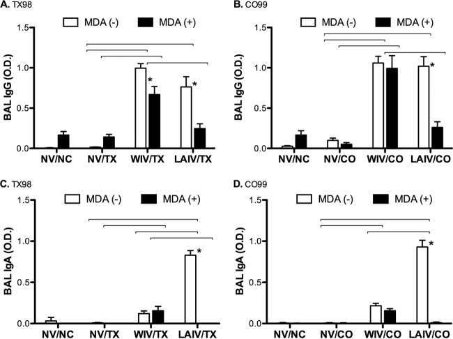
Fig 2.
Antibody levels in bronchoalveolar lavage fluid at 5 days postinfection. Mean ODs in whole-virus ELISAs are shown for IgGs against TX98 antigen (A) and CO99 antigen (B) and for IgAs against TX98 antigen (C) and CO99 antigen (D). Groups challenged with TX98 are represented in panels A and C, whereas groups challenged with CO99 are represented in panels B and D. Open bars designate groups without MDA, and solid bars designate groups with MDA. Statistically significant differences between MDA statuses within a vaccine group are marked with asterisks, and differences between vaccine treatment groups with matched MDA statuses and challenge virus strains are identified by connecting lines (P < 0.05).

