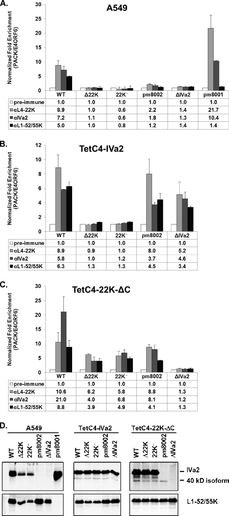
Fig 5.
ChIP assays for packaging protein binding to the PS in vivo. A549 (A), TetC4-IVa2 (B), and TetC4-22K-ΔC (C) cells were infected with Ad5-WT, Δ22K, 22K−, pm8002, ΔIVa2, or pm8001 viruses, and L4-22K-, IVa2-, and L1-52/55K-specific antibodies were used for ChIP analysis to quantify binding to the PS. The readout is presented as normalized fold enrichment as calculated by dividing the number of copies of PS pulled down specifically by the antibody-antigen complex by the number of copies of the E4-ORF6 fragment pulled down nonspecifically by the antibody. For each individual virus infection, the fold enrichment value of each antibody was normalized to that of the preimmune serum negative control. (D) Western blot analysis of ChIP input samples for determination of IVa2 and L1-52/55K protein expression levels.

