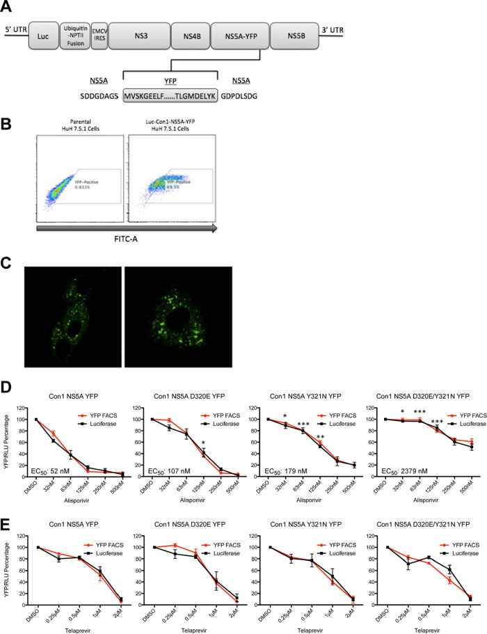
Fig 2.
(A) Schematic diagram of the NS5A-YFP reporter subgenomic Con1 replicon. NPTII, neomycin phosphotransferase gene; EMCV IRES, encephalomyocarditis virus internal ribosomal entry site. (B) The YFP-positive cell population was enriched using the BD FACSAria sorter. (C) Con1-NS5A-YFP Huh-7.5.1 cells were visualized via confocal analysis for 5 days. (D) Con1-WT NS5A-YFP, Con1-WT D320E NS5A-YFP, Y321N Con1-WT, and D320E/Y321N NS5A-YFP Huh-7.5.1 cells (500,000) were exposed to increasing concentrations of alisporivir and analyzed for YFP content 3 days after drug treatment. The percentage of YFP-positive cells treated with the DMSO control was arbitrarily fixed at 100. Results are representative of 3 independent experiments. Error bars represent standard errors from triplicates. (E) Same as panel D, except that luciferase activity in cell lysates was quantified; the percentage of luciferase activity in cells treated with the DMSO control was arbitrarily fixed at 100. Error bars represent standard errors from triplicates. Statistical significance was measured between each mutant construct in relation to Con1-WT NS5A-YFP for the following drug concentrations: 32, 63, and 125 nM. *, P < 0.05; **, P < 0.01; ***, P < 0.001.

