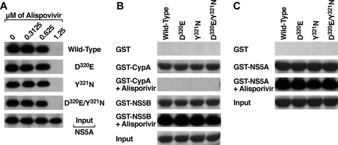
Fig 3.
(A) GST-CypA (10 μg) was mixed with wild-type, D320E, Y321N, and D320E/Y321N NS5A Con1-His proteins (200 ng) for 3 h at 4°C in the presence or absence of increasing concentrations of alisporivir. Glutathione beads were added to the GST-CypA–NS5A mixture for 30 min at 4°C and washed. Bound material was eluted and analyzed by Western blotting using anti-His antibodies. Results are representative of two independent experiments. (B) Same as panel A, except that GST, GST-CypA, or GST-NS5B (10 μg) was mixed with wild-type, D320E, Y321N, and D320E/Y321N NS5A Con1-His proteins (200 ng) for 3 h at 4°C in the presence or absence of 1.25 μM alisporivir. (C) Same as panel A, except that GST or GST-NSS5A (10 μg) was mixed with wild-type, D320E, Y321N, and D320E/Y321N NS5A Con1-His proteins (200 ng) for 3 h at 4°C in the presence or absence of 1.25 μM alisporivir.

