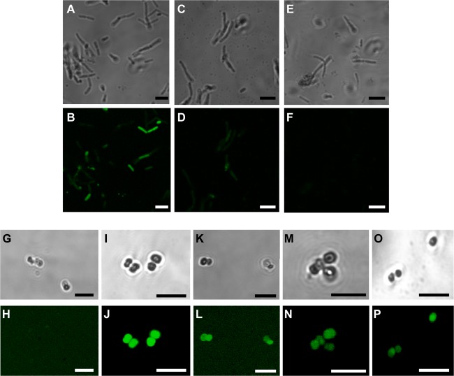
Fig 1.
Confocal laser scanning microscopy images of E. coli BL21AI (A to F) and M. luteus 10240 (G to P) incubated with fluorescein-labeled peptides (30 μmol/liter). Phase contrast (top rows) and fluorescence images (bottom rows) were taken of E. coli BL21AI cells incubated with 5(6)-carboxyfluorescein (Cf)-oncocin (A, B) and Cf-penetratin-oncocin (C, D) for 50 min and with the Cf-penetratin-Cys dimer for 90 min (E, F) or M. luteus 10240 cells incubated with Cf-pyrrhocoricin (G, H), Cf-penetratin-pyrrhocoricin (I, J), Cf-drosocin (K, L), CF-penetratin-drosocin (M, N), and the Cf-penetratin-Cys dimer (O, P). TAMRA was added to quench the fluorescence background of the medium. Bars equate to 5 μm.

