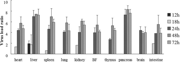
Fig 3.

Virus titer of DHAV-C in the internal organs of experimentally infected ducklings. Seven organs were obtained from the experimentally infected ducklings, and viral loads were quantified by the real-time PCR at 1 to 72 h postinfection. One of the spleen samples at 18 h generated the lowest normalized CT values (ΔCT), and this was used to calibrate the data. The calibrated quantification ratios assessed by 2−ΔΔCT were converted into logarithmic values, and the results are shown in histograms. All histograms are arranged according to the time after infection (i.e., 12 to 72 h). The virus titer was not detected at 1 and 6 h postinfection.
