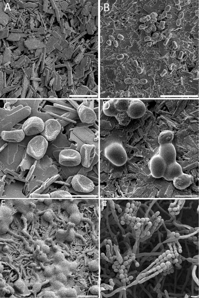
Fig 2.

Early stages in the germination of P. rubens. Conidia were applied in a dry state to gypsum with additional nutrients and incubated at 97% RH and 21°C. (A) Control: gypsum with additional nutrients inoculated with SDW. No alterations of the gypsum surface are visible after 55 h of incubation. The gypsum substrate is visible as sharp and square crystals; this is distinct from panel E, where the conidia and ECM appear smooth and round. (B) After inoculation, conidia appear dehydrated on the surface (t = 5 min). (C) Conidia remain dehydrated on the surface (t = 1 h). (D) ECM is visible on the conidia, and conidia attain an ellipsoidal shape (t = 7 h). (E) Germination tubes are formed, ECM increases with time, and ECM-covered conidia are clustered (t = 26 h). (F) Good germination and development to conidiophores, abundant aerial growth, and conidium formation are visible on the gypsum samples (t = 97 h). Scale bars: 5 μm (C, D, E, F), 10 μm (A), and 20 μm (B). The pictures shown represent specific growth phases and were not necessarily taken at the onset of the growth phase. For the onset, see Fig. 1.
