Fig 3.
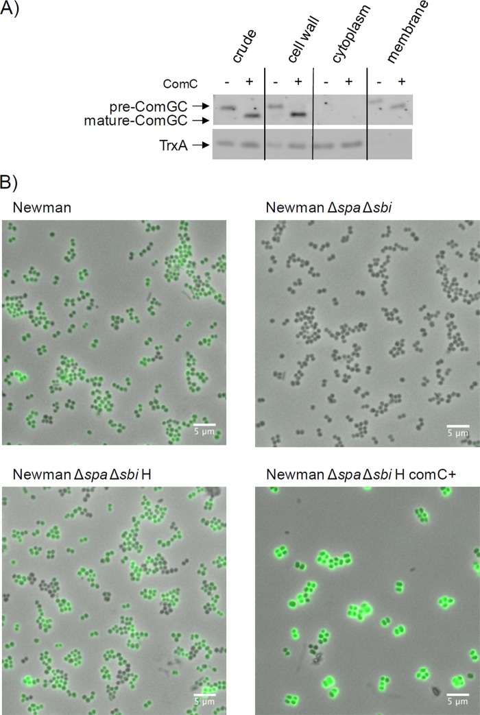
ComGC localizes to the membrane, cell wall, and cell surface of S. aureus. (A) To determine the subcellular localization of ComGC in S. aureus RN4220/pRIT:sigH or S. aureus RN4220/pRIT:sigH containing pCN51:comC, the cells were grown in LB broth for 5 h, collected by centrifugation, and incubated for 1 h at 37°C in protoplast buffer (50 mM Tris-HCl [pH 7.6], 0.145 M NaCl, 30% sucrose, 0.01% DNase, and EDTA-free Complete protease inhibitors [Roche]). The cell wall fraction (i.e., protoplast supernatant) was obtained by centrifugation (20 min, 3,000 × g, 4°C). Protoplasts were disrupted by osmotic shock in 0.05 M Tris-HCl (pH 7.6) with 30 min of incubation on ice and vortexing at 5-min intervals. Cytosolic and solubilized membrane proteins were collected as previously described (24). SDS-PAGE and Western blotting with specific antibodies against ComGC or the cytoplasmic control protein TrxA were performed as described in the legend to Fig. 1. (B) Cell surface exposure of ComGC was assessed in S. aureus Newman Δspa Δsbi or the parental strain (Newman) by immunofluorescence microcopy. For this purpose, cells were grown for 5 h in LB broth, and 1 unit of cells (by optical density at 600 nm) was collected by centrifugation (8,000 rpm, 5 min, 4°C). The cell pellet was resuspended in phosphate-buffered saline-Tween 20 (PBST) plus 2% bovine serum albumin (BSA) and incubated for 10 min on ice. Next, the cells were incubated for 60 min with ComGC-specific polyclonal rabbit antibodies (1:400 in PBST plus 1% BSA). Unbound antibodies were removed by three washes in PBST, and cell-bound ComGC antibodies were visualized using goat-anti-rabbit Alexa Fluor 488 antibodies (Life Technologies) and a Leica DM5500 B microscope. The overlay of phase-contrast and fluorescence microscopy images was done with imageJ. The strains containing pRIT:sigH for σH production are indicated by “H”; the strain containing pCN51:comC for ComC production is indicated by “ComC+.” The magnification is indicated by scale bars.

