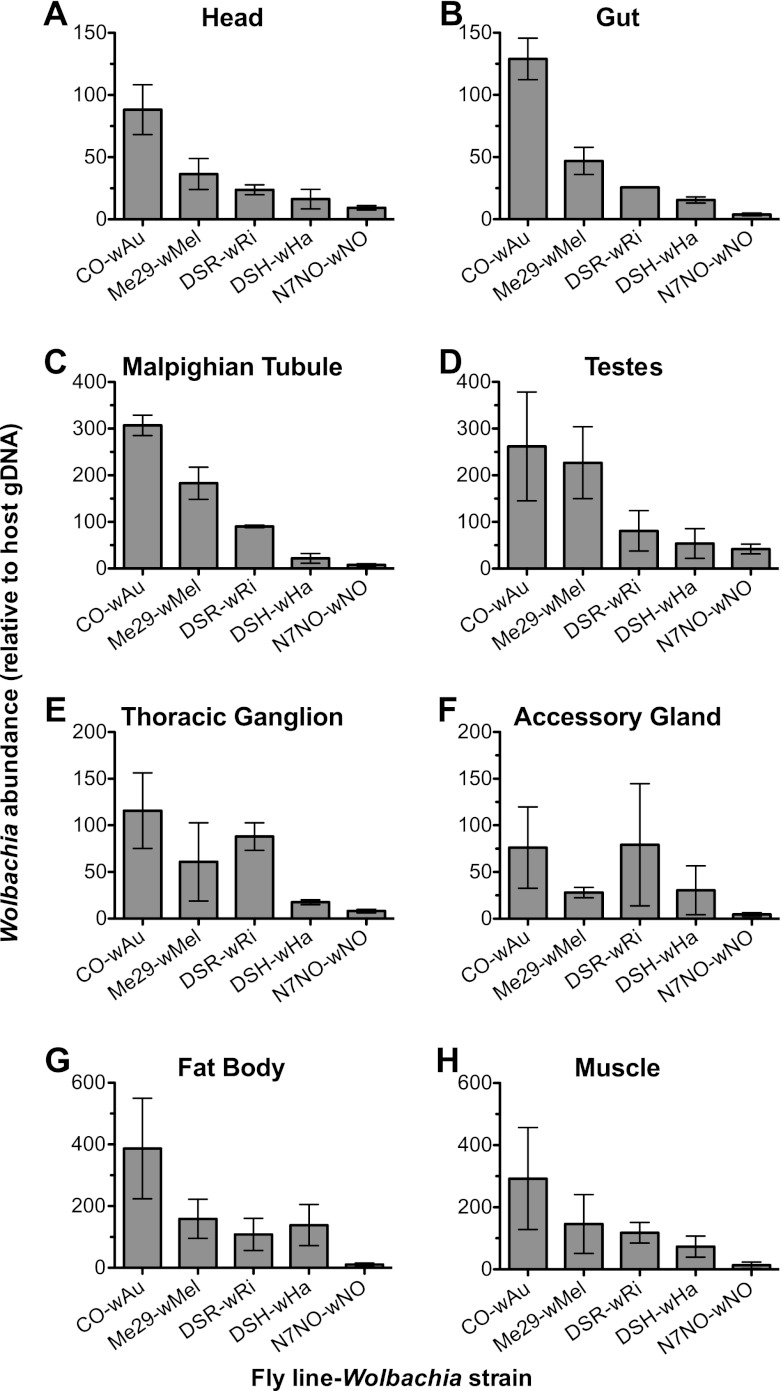
Fig 1.
Relative Wolbachia abundance in tissues isolated from D. simulans flies. The graphs show the Wolbachia abundance relative to a host reference gene (Actin 5C) in dissected tissues as determined by qPCR. Tissues are the head (A), gut (B), Malpighian tubules (C), testes (D), thoracic ganglion (E), accessory glands (F), fat body (G), and muscle (H). Dissected tissues were pooled from five individual flies, and the experiment was repeated across three independent fly cohorts. The error bars represent standard errors. P values were interpreted as significant when <0.01. A correlation between the Wolbachia density within a tissue and the mediation of antiviral protection was observed with the head (P = 0.0052), gut (P = 0.0001), and Malpighian tubules (P = 0.0001) but not in the testes (P = 0.0430), thoracic ganglia (P = 0.0117), accessory glands (P = 0.2279), fat body (P = 0.1005), or muscle (P = 0.1055).

