
Fig 2.
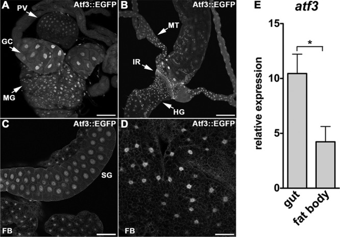
Atf3 is expressed in tissues central to metabolic and immune homeostasis. (A to D) Anti-GFP antibody detected a nuclear Atf3::EGFP signal in confocal micrographs of the gut (A and B), salivary glands (C), and fat body (C and D) dissected from third-instar atf3[gBAC]/+ larvae. GC, gastric ceca; MG, midgut; PV, proventriculus; HG, hindgut; IR, imaginal ring; MT, malpighian tubules; SG, salivary glands; FB, fat body. Scale bars are 100 μm (A to C), and 50 μm (D). (E) atf3 mRNA expression in the larval gut is about 2.5-fold higher than in the fat body as determined by qRT-PCR. The data are means and SEM; *, P < 0.05.