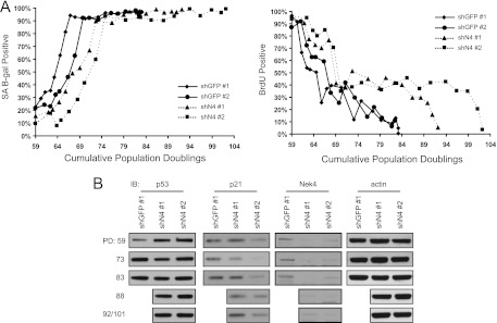
Fig 6.
Characterization of extended-life-span NEK4-suppressed cells. (A) (Left) SA β-Gal staining. BJ shGFP no. 1 and no. 2 and BJ shN4 no. 1 and no. 2 cells were fixed and stained with X-Gal to identify the percentages of cells with SA β-Gal activity at the indicated population doublings. At least 100 cells were scored for each time point. (Right) Parallel BrdU incorporation assays. Cells were also incubated with BrdU for 48 h to assess the percentages of proliferating cells at the indicated population doublings. At least 100 cells were scored for each time point. (B) Immunoblotting (IB) of markers of senescence. Steady-state levels of p53, p21, Nek4, and actin were assessed via immunoblotting in BJ shGFP no. 1 or BJ shN4 no. 1 and no. 2 cells at the indicated PD. Control cells senesced prior to PD 88, as can be seen in panel A. The last time points for shN4 no. 1 and no. 2, PD 92 and 101, respectively, were assessed together in the bottom row.

