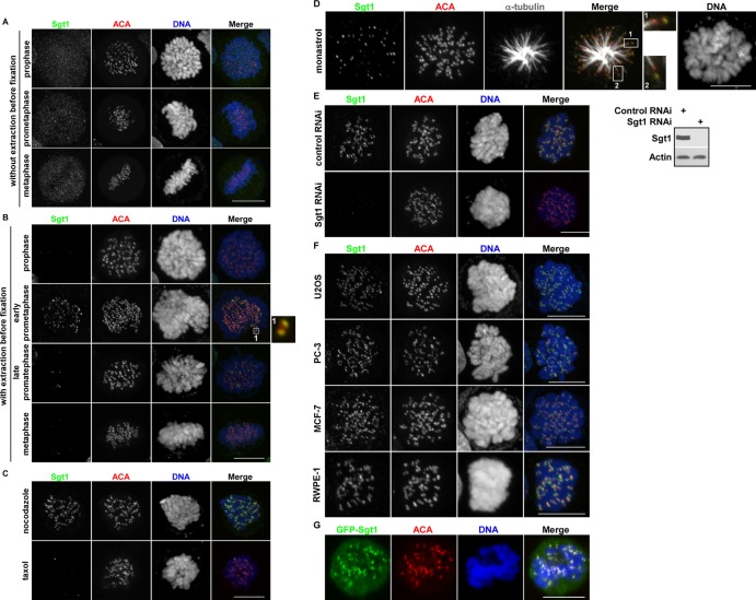
Fig 1.
Sgt1 dynamically localizes at the kinetochore during mitosis. (A and B) HeLa cells in various mitotic stages were directly fixed in 4% paraformaldehyde (A) or extracted by 1% Triton X-100 treatment (B) and then stained for IF with anti-Sgt1 antibody or ACA and with 4′,6-diamidino-2-phenylindole (DAPI). Individual and merged fluorescent images are presented. An enlarged image on the right shows that Sgt1 (green) colocalizes with the centromere (red). Bar, 10 μm. (C) HeLa cells were treated with 1 μM nocodazole or 1 μM paclitaxel for 2 h and then subjected to IF staining as described for panel B. Bar, 10 μm. (D) HeLa cells were treated with 68 μM monastrol for 2 h and processed for IF staining with anti-Sgt1, ACA, or anti-α-tubulin antibodies and then DAPI. Two enlarged images (boxes 1 and 2) on the right of the merged image show that single kinetochores with microtubule attachment do not show Sgt1 staining, whereas other kinetochores lacking microtubule attachment show positive Sgt1 staining. Bar, 10 μm. (E) HeLa cells were transfected with control or Sgt1 siRNAs for 72 h and subjected to immunoblotting (right panel) or IF staining (left panel) with the indicated antibodies. Nocodazole was added 2 h before the cells were harvested for IF staining. Bar, 10 μm. (F) U2OS, PC-3, MCF-7, and RWPE-1 cells were treated as described for panel B. Nocodazole was added to these cells before they were subjected to anti-Sgt1 IF staining. Bar, 10 μm. (G) U2OS cells were transfected with GFP-Sgt1 and selected by use of G418 for 2 weeks. Cells were treated with nocodazole and subjected to IF staining with anti-GFP antibody and ACA. Bar, 10 μm.

