
Fig 6.
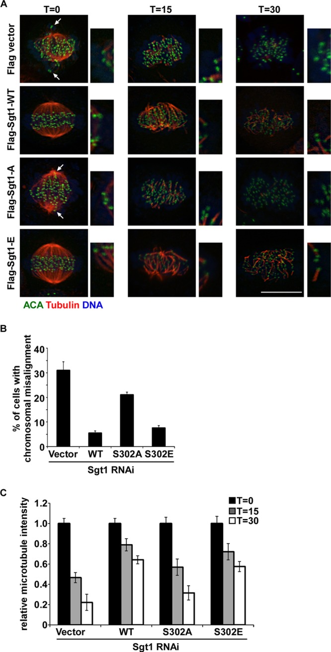
Phosphorylation of Sgt1 contributes to stable kinetochore-microtubule attachments. (A) After HeLa cells were cotransfected with the indicated murine Flag-Sgt1 constructs and Sgt1 siRNA as described for Fig. 3B, cells were treated with monastrol for 8 h and released for 1 h into fresh medium in the presence of MG132, resulting in a metaphase-arrested population. Cells were then incubated for the indicated times at 4°C, fixed, and processed for IF staining with antibodies against α-tubulin or centromere (ACA). Arrows indicate misaligned chromosomes with kinetochore staining not on the metaphase plate. Enlarged representative kinetochore-microtubule connections are shown on the right. Bar, 10 μm. (B) The cells in panel A were synchronized by thymidine block for 24 h and released into fresh medium for 8 h, followed by 1 h of incubation with MG132, resulting in a metaphase-arrested population. Percentages of cells with misaligned chromosomes were quantified. Results represent the averages of three independent experiments (means ± standard errors; n > 100 cells/condition). (C) Quantification of microtubule density for the cells shown in panel A. Average microtubule intensity (mean ± standard error; n = 5 cells) was measured by using ImageJ under each condition. Intensities are expressed relative to total cellular areas and normalized against time zero (T=0) for each condition (microtubule intensity at time zero was set as 100%).