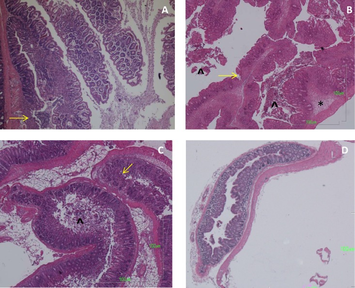
Fig 6.
H&E-stained colons from mice at 7 days postinfection. (A) Inflammatory polyps in the proximal colon of a mouse infected with WT S. Typhimurium 14028s. There is an inflammatory cell infiltrate in each of the polyps and an area of mucosal ulceration on the lower polyp. (B) Rectum from a mouse infected with WT S. Typhimurium 14028s, with submucosal edema (asterisk), sloughed epithelial cells in the lumen (arrowhead), and inflammatory infiltrates in the mucosa. (C) Rectum from a mouse with a sifA mutant infection, showing intense inflammation, a lesser degree of submucosal edema, mucosal ulcerations, and epithelial cells in the lumen. (D) Rectum from a mouse infected with the ssaV mutant; the tissue is normal. Yellow arrows point to areas of mucosal erosions. Magnification for all photos, ×40.

