
Fig 3.
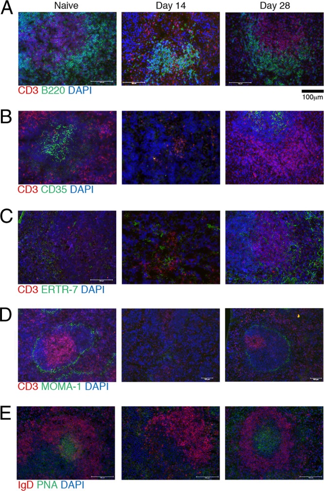
Acute infection results in the disorganization of splenic structures. (A to E) Splenic sections were taken from infected mice at the indicated time points and stained for immunofluorescent analysis. Structures were evaluated by staining for T and B cell zones with CD3 (red), B220 (green), and DAPI (blue) (A); follicular DCs with CD3 (red), CD35 (green), and DAPI (blue) (B); fibroblast reticular cells with CD3 (red), ERTR-9 (green), and DAPI (blue) (C); metallophillic macrophages using CD3 (red), MOMA-1 (green), and DAPI (blue) (D); or GCs using IgD (red), PNA (green), and DAPI (blue) (E).