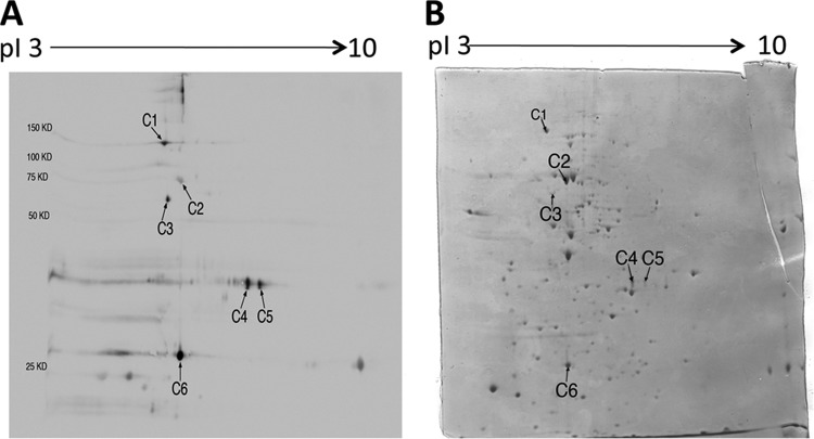
Fig 1.
2-DE immunoblot showing T. gondii antigens recognized by IgG antibodies in sera of infected CBA/J mice treated with sulfadiazine to inhibit proliferation of tachyzoites during the chronic stage of infection (A) and a Coomassie-stained 2-DE gel indicating protein spots corresponding to each of the antigens recognized by the IgG antibodies (B). Tachyzoites (1 × 10⁸ parasite) were solubilized in sample buffer and subjected to 2-DE. The gel was stained with Coomassie brilliant blue and scanned with an HP Scanjet G4050. The tachyzoite antigens separated by 2-DE were transferred to a nitrocellulose membrane, and the membrane was subjected to immunoblotting with the pooled sera from the infected and sulfadiazine-treated mice (see Materials and Methods). The identity of each antigen indicated (C1 to C6) on the immunoblot is described in Table 1.

