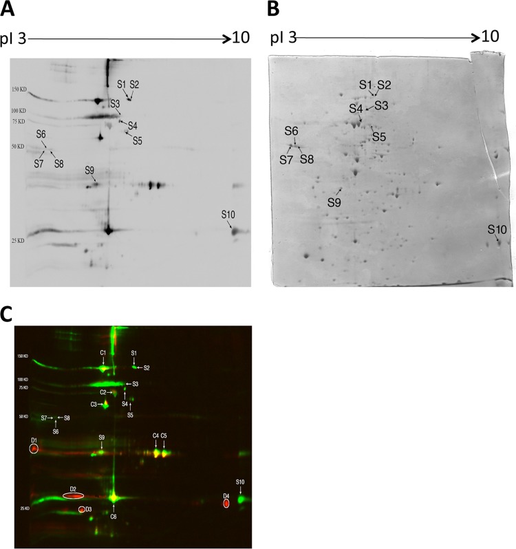
Fig 3.
2-DE immunoblot showing T. gondii antigens recognized by IgG antibodies in sera of CBA/J mice with cerebral proliferation of tachyzoites during the chronic stage of infection (A) and a Coomassie-stained 2-DE gel indicating protein spots corresponding to each of the antigens recognized by the IgG antibodies (B). Tachyzoites (1 × 10⁸ parasite) were solubilized in sample buffer and subjected to 2-DE. The gel was stained with Coomassie brilliant blue and scanned with an HP Scanjet G4050. The tachyzoite antigens separated by 2-DE were transferred to a nitrocellulose membrane, and the membrane was subjected to immunoblotting with the pooled sera from the infected mice (see Materials and Methods). The spots indicated (S1 to S10) are those recognized only by or mostly by the IgG antibodies of mice with cerebral proliferation of tachyzoites compared to the immunoblot with the IgG antibodies of mice treated with sulfadiazine to inhibit tachyzoite growth during the chronic stage of infection shown in Fig. 1A. The identity of each antigen indicated on the immunoblot is described in Table 3. Panel C is an overlay of the blots shown in Fig. 1A and 3A using two different colors: red for the spots shown in Fig. 1A and green for the spots shown in Fig. 3A. Whereas the spots corresponding to C1 to C6 are yellow, the spots corresponding to S1 to S10 are exclusively green. There are multiple spots shown exclusively red (D1 to D4), indicating that these antigens are recognized only by or mostly by IgG antibodies from mice that did not have active tachyzoite proliferation.

