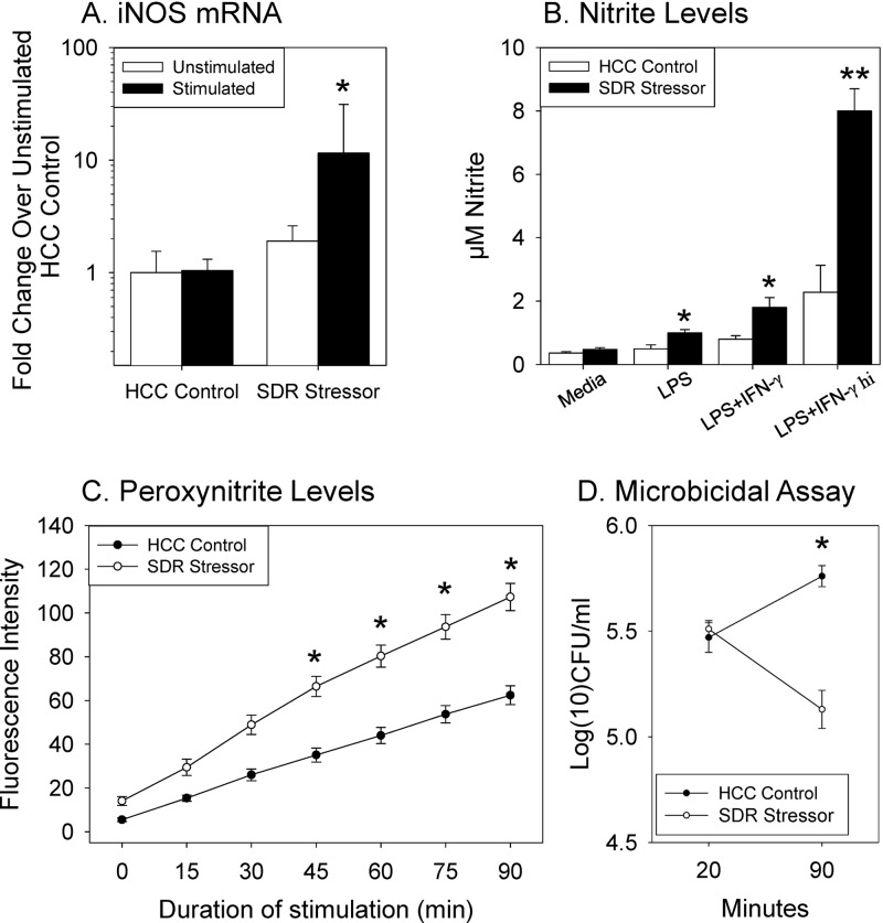
Fig 1.
Exposure to the SDR stressor significantly enhances splenic macrophage activity. (A) Significant increase in iNOS mRNA expression in SDR mice stimulated with E. coli compared to mRNA in nonstressed HCC control mice. Values are means ± the strandard errors (SE) (n = 9 SDR and 8 HCC from three different experiments; *, P < 0.05 versus HCC E. coli). (B) Significant increase in the levels of nitric oxide-derived nitrite by LPS-, LPS + IFN-γ (5 ng/ml)-, and LPS + IFN-γ hi (10 ng/ml)-stimulated splenic macrophages from mice exposed to SDR compared to nonstressed HCC control mice. Values are means ± the SE (n = 8 SDR and n = 8 HCC from two different experiments; *, P < 0.05 versus cells from HCC controls; **, P < 0.05 versus all other groups). (C) Significant increase in production of peroxynitrite in PMA/LPS/IFN-γ-stimulated splenic macrophages from mice exposed to the SDR stressor compared to nonstressed HCC mice. Values are fluorescence intensity means ± the SE (n = 15 SDR and n = 18 HCC from five different experiments; *, P < 0.001). (D) Significant decrease in E. coli bacterial counts in SDR splenocytes at 90 min. Bacterial counts determined via plate counts obtained 20 min after addition of bacteria (to calculate number of bacteria phagocytosed) and 90 min after addition of bacteria (to calculate number of remaining phagocytosed bacteria). Values are means ± the SE (n = 13 SDR and n = 13 HCC from three different experiments; *, P < 0.05 versus SDR 20 min).

