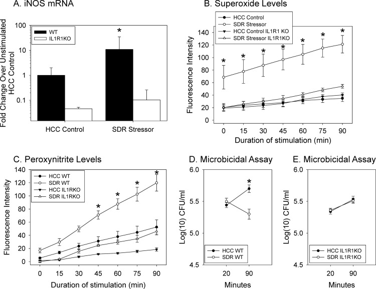
Fig 5.
Stressor-induced increases in splenic macrophage activity do not manifest in IL-1R1−/− mice. (A) Exposure to the SDR stressor did not enhance iNOS mRNA in the splenic macrophages from IL-1R1−/− mice stimulated with E. coli for 90 min. The stressor did increase iNOS mRNA in wild-type mice (*, P < 0.05; n = 8 WT HCC, n = 7 WT SDR, n = 3 IL-1R1−/− HCC, and n = 3 IL-1R1−/− SDR from two different experiments). (B) Superoxide production did not differ with stress in IL-1R1−/− mice (*, P < 0.05, WT SDR versus all other groups; n = 7 WT HCC, n = 5 IL-1R1−/− HCC, n = 7 WT SDR, and n = 5 IL-1R1−/− SDR from two different experiments). (C) The production of peroxynitrite by PMA/LPS/IFN-γ-stimulated splenic macrophages from WT mice exposed to the SDR stressor was significantly higher than peroxynitrite production by all other groups (*, P < 0.05 versus all other groups at designated time points). Peroxynitrite production by splenic macrophages from IL-1R1−/− mice exposed to the SDR stressor was significantly lower than peroxynitrite production by splenic macrophages from the WT mice exposed to the SDR stressor (*, P < 0.05 at 45, 60, 75, and 90 min; n = 6 WT HCC, n = 6 WT SDR, n = 5 IL-1R1−/−, and n = 5 IL-1R1−/− from two different experiments). (D) The number of E. coli killed in cultures containing splenic macrophages from WT mice exposed to the SDR stressor was significantly different at the 90-min time point (*, P < 0.05 versus HCC control). (E) However, there was no difference in the number of bacteria killed by the splenic macrophages from IL-1R1−/− mice exposed to the SDR stressor compared to the nonstressed HCC IL-1R1−/− mice (n = 8 WT HCC, n = 8 WT SDR, n = 14 IL-1R1−/− HCC, and n = 15 IL-1R1−/− SDR from three different experiments).

