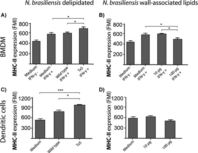
Fig 6.
(A) Bar graphs showing the expression of MHC-II molecules in IFN-γ-activated bone marrow-derived macrophages (BMDM) infected with wild-type or Tx5 N. brasiliensis. As controls, BMDM were incubated in medium with (medium IFN-γ+) or without (medium IFN-γ−) IFN-γ, and expression of MHC-II molecules was assessed by flow cytometry. (B) Expression of MHC-II molecules in IFN-γ-activated BMDM stimulated with several concentrations of N. brasiliensis wall-associated lipids. As controls, BMDM were incubated in medium with (medium IFN-γ+) or without (medium IFN-γ−) IFN-γ, and expression of MHC-II molecules was assessed by flow cytometry. (C) MHC-II molecules in dendritic cells infected with wild-type or Tx5 N. brasiliensis. As controls, DCs were incubated in medium alone, and expression of MHC-II molecules was assessed by flow cytometry. (D) Expression of MHC-II molecules in DCs stimulated with several concentrations of N. brasiliensis wall-associated lipids. As controls, DCs were incubated in medium alone, and expression of MHC-II molecules was assessed by flow cytometry. Data expressed as means ± standard deviations of the fluorescence mean intensity (FMI) of MHC-II expression. *, P < 0.05; ***, P < 0.0001 as assessed using a Bonferroni posttest.

