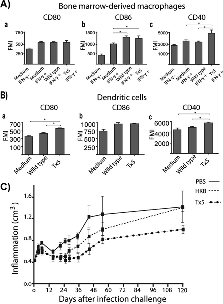
Fig 7.
(A) Bar graphs showing the expression of several T cell costimulatory molecules in IFN-γ-activated BMDM infected with wild-type or Tx5 N. brasiliensis. As controls, BMDM were incubated in medium with (medium IFN-γ+) or without (medium IFN-γ−) IFN-γ, and expression of MHC-II molecules was assessed by flow cytometry. (B) Expression of several T cell costimulatory molecules in dendritic cells infected with wild-type or Tx5 N. brasiliensis. As controls, DCs were incubated in medium alone, and expression of MHC-II molecules was assessed by flow cytometry. Data expressed as means ± standard deviations of the fluorescence mean intensity (FMI) of MHC-II expression. *, P < 0.05; ***, P < 0.0001 as assessed using a Bonferroni posttest. (C) Dot plot showing the level of footpad inflammation (in cm3) in mice immunized with heat-killed N. brasiliensis (HKB), PBS, or Tx5 N. brasiliensis and challenged with wild-type N. brasiliensis 15 days after immunization.

