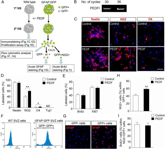
Figure 1.
Characterization of control and PEDF-treated neurospheres derived from the SVZ of adult wild-type or GFAP:GFP transgenic mice. A, Experimental flow chart. Secondary neurospheres derived from the SVZ of either adult wild-type or GFAP:GFP transgenic mice were grown in the absence or presence of PEDF (50 ng/ml) for 5 d, and then assayed as indicated. B, RT-PCR analysis for expression of PEDF receptor in SVZ neurospheres. C, D, Wild-type control and PEDF-treated secondary neurosphere cells were immunostained for nestin, NG2, O4, or Tuj1. Immunostaining images (C) and quantification data (D) showing that PEDF significantly increased the numbers of NG2+ and O4+ cells. E, Quantification of BrdU incorporation and Ki67 labeling in wild-type control and PEDF-treated secondary neurospheres. PEDF did not alter proliferation of neurosphere cells. F, FACS plots for GFP+ and GFP− cell fractions dissociated from adult SVZ GFAP:GFP neurospheres. GFP+ selection was set using wild-type SVZ neurospheres as a negative control. G, GFAP immunostaining acutely done on plated GFP+ and GFP− cells FACS-purified from GFAP:GFP neurospheres. H, Graphic representation of flow cytometric quantification of percentages of NG2+ cells among GFP+ cells in control and PEDF-treated secondary GFAP:GFP neurospheres, showing that PEDF promoted NG2 induction in GFP+ cells. I, Quantification of BrdU incorporation among GFP+NG2+ cells FACS-purified from control and PEDF-treated secondary GFAP:GFP neurospheres, indicating that PEDF-mediated NG2 induction in GFAP:GFP+ precursors was not due to selective proliferation of GFP+NG2+ cells. ND, Not determined. Results are means ± SEM of three or four independent experiments. *p < 0.05, **p < 0.01, Student's t test. Scale bar, 30 μm.

