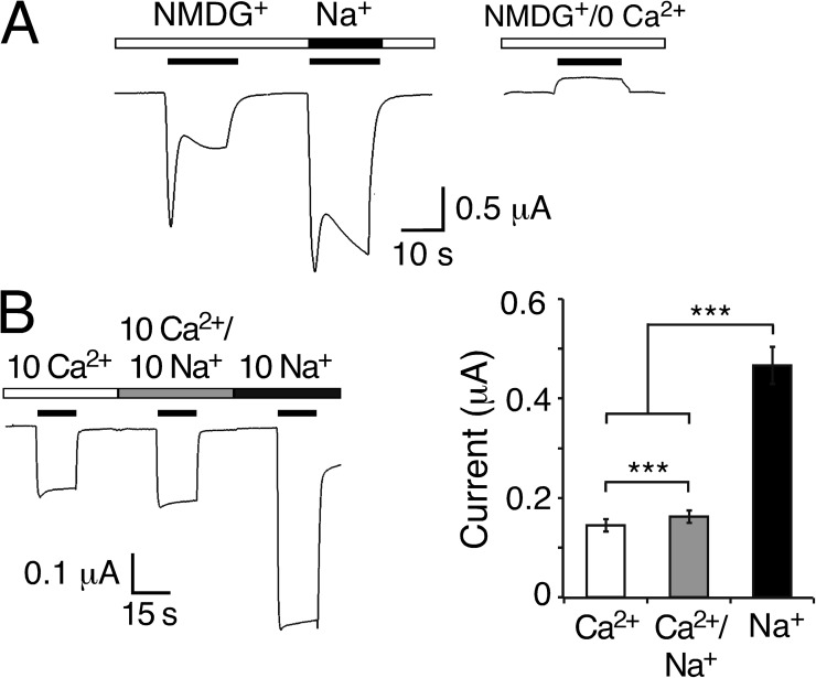
Figure 3.
Peptide-activated currents in the absence of Na+e. (A; left) Representative current trace for HyNaC2/3/5 in solutions containing 1 mM Ca2+ together with 140 mM of either NMDG+ or Na+. 1 µM RFamide I was used for activation. (Right) Control experiment in NMDG+ solution nominally free of Ca2+. Similar results were observed in four out of four oocytes. (B; left) Representative current trace for HyNaC2/3/5 in solutions containing 10 mM Ca2+, 10 mM Ca2+ and 10 mM Na+, or 10 mM Na+ (nominally free of Ca2+). Solutions additionally contained 10 mM HEPES, pH 7.4, and NMDG+ at concentrations to reach similar osmolarity (10 Ca2+: 125 mM NMDGCl; 10 Ca2+/10 Na+: 115 mM NMDGCl; and 10 Na+: 130 mM NMDGCl). Oocytes had been injected with EGTA; 2 µM RFamide I was used for activation. Holding potential was −85 mV. (Right) Quantitative comparison of current amplitudes. ***, P < 0.001.

