
Fig. 1.
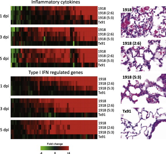
Activation of inflammatory and type I IFN-regulated genes in 1918 influenza virus-infected mouse lung tissue. Gene expression profiles in the lungs of mice infected with equivalent doses the reconstructed 1918 virus (r1918), a chimeric virus expressing either the 1918 HA and NA genes (2:6) or the 1918 HA, NA, M, NP and NS genes (5:3) compared to a contemporary H1N1 human-adapted influenza virus A/Texas/36/91 (Tx91). (Top panel) expression of inflammatory cytokine genes, (bottom panel) expression of type I IFN-regulated genes. For each infection point, the data presented are the error-weighted average expression changes calculated from four technical replicate arrays performed on three individual mice (n = 12 total). Genes shown in red were up-regulated and genes shown in green were down-regulated in infected relative to mock-infected mouse lung. At right is shown the lung pathology at 3 days post-infection. Modified from Kash et al. (2006c) (data available at http://viromics.washington.edu/publications.html).