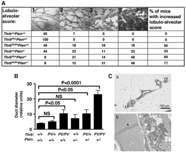
Figure 1.
Increased lobulo-alveolar development in the mammary glands of knock-in mice expressing TRβPV. (A) Mammary gland phenotypes of mice of the six genotypes. Whole mammary glands were stained with carmine red to visualize the structure of the ducts and alveolae. Four different lobulo-alveolar stages, scored from 1 to 4, could be distinguished as described in Results. The values in the table represent the percentage of mice of each genotype that could be related to one of the stages. The last column shows the percentage of animals with lobulo-alveolar development at stages 3 and 4. (B) Duct diameter in the six different genotypes. Whole mounts were photographed, and duct diameter was measured using NIH IMAGE software (ImageJ 1.34s; Wayne Rashband, NIH) (http:// rsb.info.nih.gov/ij). Shown is the average of the diameter sizes taken at four different locations on the largest duct. (C) Aberrant lobulo-alveolar development is associated with lactational changes. Hematoxylin and eosin mammary tissue sections of a wild-type (a) and a Thrbpv/+Pten+/− (b) mice. Note the dramatic dilatation of the duct (*) in the Thrbpv/+Pten+/− mouse.

