Figure 3.
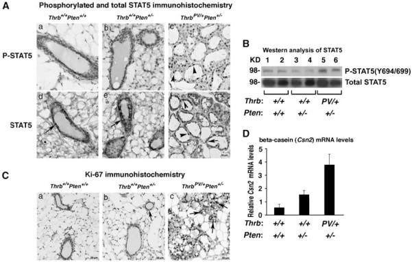
The TRβPV mutation aberrantly activates STAT5 signaling to increase cell proliferation. (A) Immunohistochemistry of P-STAT5 on mammary tissue sections in Thrb+/+Pten+/+, Thrb+/+Pten+/− and Thrbpv/+Pten+/− mice. Tissue sections were counterstained with hematoxylin (blue). Thrb+/+Pten+/+and Thrb+/+Pten+/− mammary glands are devoid of P-STAT5 positively stained cells, whereas numerous P-STAT5 positively stained cells (brown nuclei) are present in the hyperplastic Thrbpv/+Pten+/−mammary gland. (B) Western analysis of P-STAT5 (upper panel) and total STAT5 (lower panel) on mammary tissue lysates from Thrb+/+Pten+/+ (lanes 1 and 2), Thrb+/+Pten+/− (lanes 3 and 4) and Thrbpv/+Pten+/− (lanes 5 and 6) mice. Total lysates (50 μg) were analyzed as described in Materials and methods. The lanes are marked (n 1/4 2 per genotype). (C) Assessment of cell proliferation byKi-67 immunohistochemistry on mammary tissue sections in Thrb+/+Pten+/+, Thrb+/+Pten+/− and Thrbpv/+Pten+/− mice. Tissue sections were counterstained with hematoxylin (blue). (D) Levels of Csn2 encoding β-casein in the mammary glands of Thrb+/+Pten+/+,Thrb+/+Pten+/− and Thrbpv/+Pten+/− mice. Shown are Csn2 mRNA levels determined by real-time reverse transcriptase–PCR and normalized to Gapdh (glyceraldehyde-3-phosphate dehydrogenase) mRNA levels.

