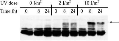
Figure 5.
Time course of RPA phosphorylation after irradiation of XP12BE cells (XP complementation group A). XPA cells were either mock-irradiated or treated with the indicated doses of UVC. At the indicated times after treatment, cell lysates were prepared and the RPA-p34 phosphorylation pattern was analyzed by gel electrophoresis, followed by immunoblotting. The arrow to the right indicates the major hyperphosphorylated form of RPA-p34.

