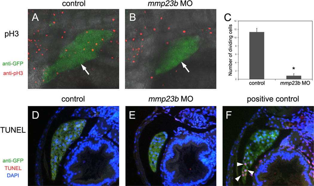
Figure 3. Cell division defect in mmp23b morphants.
(A and B) Anti-phospho Histone H3 (pH3) staining for control and mmp23b morphant embryos at 3dpf. Red: pH3 staining. Green: GFP staining. pH3 staining in the liver (B, white arrow) of mmp23b morphant is rarely observed. (C) Quantification of dividing cells by pH3 staining. *P<0.00001. (D–F) TUNEL analysis at 3dpf. Red: TUNEL staining. Green: GFP staining. Blue: DAPI staining. (D) Control embryo. (E) Mmp23b morphant. No apoptotic cells were observed in the developing liver of control or mmp23b morphant (L). (F) Positive control. Embryos were treated with DNaseI before TUNEL assay. Note apoptotic cells in the developing liver (white arrowheads).

