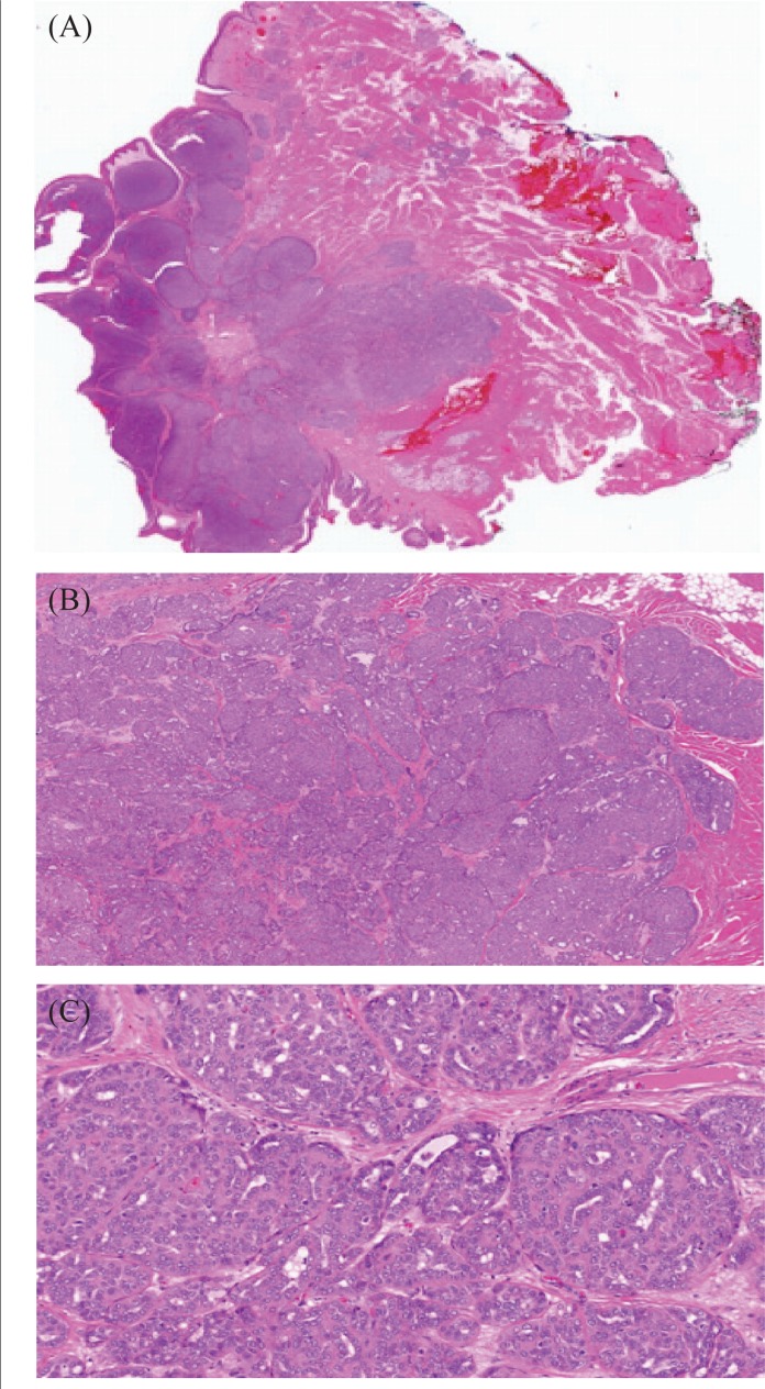
FIGURE 2.
Histology sections of adenocarcinoma not otherwise specified of minor salivary gland origin. (A) Low-power photomicrograph of the lesion, demonstrating its well-circumscribed nature in the submucosa. (B) Medium-power photomicrograph demonstrating arrangement of the neoplastic cells in cribriform nests, sheets, and focal tubules. (C) High-power photomicrograph of the neoplasm, demonstrating its cytologic features.

