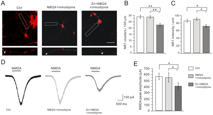
Figure 3. Zinc exposure decreases NR1 clusters and NMDA currents.
Cultured hippocampal neurons were immunostained for NR1. (A) Representative images of clustering of NR1 in control, NBQX+nimodipine and Zn+NBQX+nimodipine groups. For each image, a higher magnification shows the dendritic segment (enclosed in white box) studded with numerous clusters, indexed by white arrows. Scale bar, 50 µm. (B) and (C) show the quantification of (A). Both the number and mean intensity of NR1 clusters were significantly decreased after zinc treatment. The mean intensities were 85.4±4.0 (control; n = 27), 90.2±4.0 (NBQX+nimodipine; n = 24), and 71.5±4.0 (Zn+NBQX+nimodipine; n = 27). (D) Representative whole-cell currents in response to agonist solution containing 30 µM NMDA, 10 µM glycine, 1 µM TTX, 10 µM bicuculline, 10 µM NBQX and 1 µM strychnine in cultured pyramidal cells from control, NBQX+nimodipine and Zn+NBQX+nimodipine groups. The averaged peak amplitude of NMDA currents in Zn+NBQX+nimodipine group was reduced, as shown in (E). *, P<0.05, **, P<0.01.

