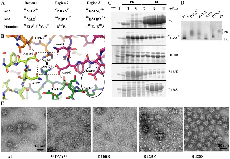
Figure 2. Analysis of dodecahedra formation by Ad3 Pb mutants.
(A) Amino acid sequence of Pb protein regions responsible for Dd integrity. (B) Structure of regions 2 and 3 involved in Dd formation. These residues are located around the 2-fold axes of the dodecahedron. The light and dark green molecules belong to one Pb, the magenta and orange molecules to another one. Dotted lines mark potential hydrogen bonds, although they cannot be identified unambiguously due to the limited resolution. (C) Sucrose density gradients of rwtDd and mutants. Cell extracts obtained from expressing HF cells were fractionated and analyzed on denaturing PAGE as described in Material and Methods. (D) Native gel analysis of Q-Sepharose fractions eluted with 370 mM NaCl and revealed with CBB stain. Pb and wt Dd were used as internal standards. Note the shift of Dd bands due to changed net charges of Dd constructs. (E) Electron microscopy of wt and mutant Dd. Samples shown in C were analyzed after dialysis.

