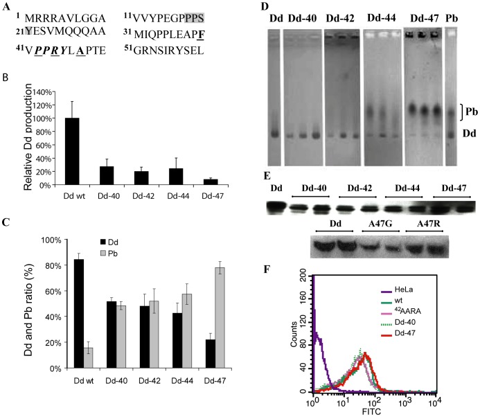
Figure 4. Analysis of Ad3 Pb mutants.
(A) Sequence of Ad3 Pb N-terminus. The first PPxY motif is underlain in grey, the second PPxY motif is written in bold italics. The first amino acid residue of deletants is written in bold and underlined. (B) Dd production in the baculovirus system. Cell lysates were purified by centrifugation on a sucrose density gradient. Fractions recovered from 15–25% sucrose (Pb) and from 30–40% sucrose (Dd) were pooled separately, run on SDS-PAGE and analyzed by gel densitometry. (C) Relative production of Dd and Pb by N-terminal deletants. Data for Dd and free Pb production are expressed as a percentage of total Pb protein expression. The average from four electrophoretic runs is shown. (D) Native agarose gel electrophoresis (CBB stain) of proteins recovered from the Q-Sepharose column at a salt concentration corresponding to the elution of Dd. The fractions with the highest amount of protein were used. (E) Internalization of Dd mutants. Q-Sepharose-purified proteins were applied onto HeLa cells and intracellular Dd was visualized by Western blot or by flow cytometry (F) as described in Materials and Methods.

