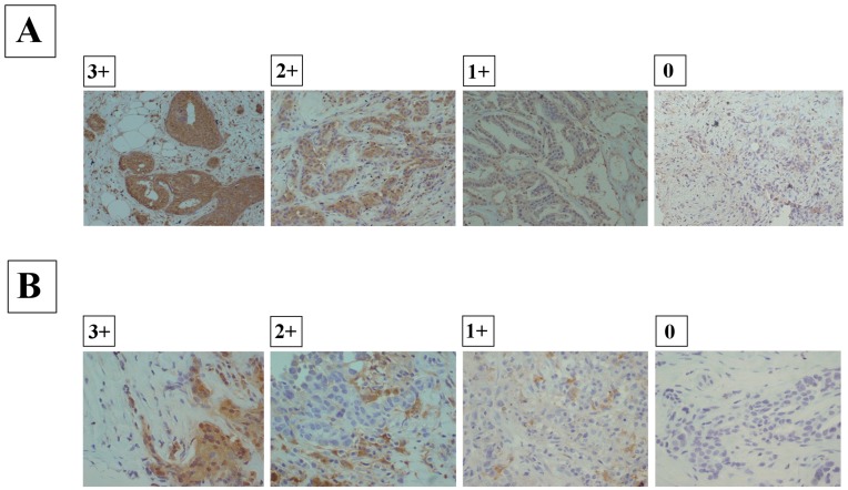
Figure 2. Immunohistochemical staining of CD133 or ALDH1.
(A), Immunohistochemical determination of CD133 expression. The CD133 antibody stained intensely at the membrane and in the cytoplasm of cancer cells. Scores were applied as follows: 0, negative staining in all cells; 1+, weakly positive or focally positive staining in <10% of cells; 2+, moderately positive-staining in 10%-50% of cells; and score 3+, strongly positive-staining, involving 50% or more of the cells. (B), Immunohistochemical determination of ALDH1 expression. The ALDH1 antibody stained intensely at the membrane and in the cytoplasm of cancer cells. Scores were applied as follows: 0, negative staining in all cells; 1+, weakly positive or focally positive staining in <10% of cells; 2+, moderately positive-staining in 10%-50% of cells; and score 3+, strongly positive-staining, involving 50% or more of the cells.

