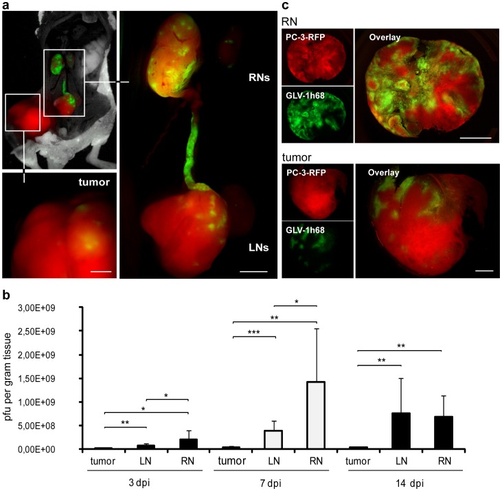
Figure 3. Colonization of lymph node metastases and PC-3-RFP tumors by GLV-1h68.
1×10⁷ pfu GLV-1h68 were i.v. injected into PC-3-RFP tumor-bearing mice. (a) PC-3-RFP tumor-bearing mouse 3 days after injection of GLV-1h68. (b) Virus titers in PC-3-RFP tumors, LNs and RNs 3, 7 and 14 days after injection of GLV-1h68. Virus injection was performed 55 days post tumor cell implantation. Tumors and lymph node metastases of 6 mice were analyzed per group (n = 6). (c) 100 µm sections of a PC-3-RFP tumor and a renal lymph node metastasis 77 days post implantation and 7 days after virus injection. PC-3-RFP cells are shown in red, by GLV-1h68 expressed GFP in green. All images are representative examples. Scale bars represent 1 mm (b) and 2 mm (c).

