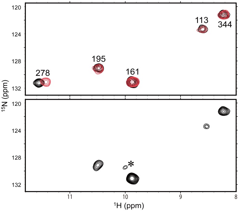
Figure 2. Probing collagen binding of Hsp47 using 1H-15N HSQC peaks originating from the tryptophan indole groups.
1H-15N HSQC spectra of the ε-imino groups of the tryptophan residues of wild-type (upper) and the W280Y mutant (lower) of Hsp47 in the absence (black) or presence (red) of trimeric collagen peptide. The assignment of the Trp280 peak was made by comparing the spectra of the wild-type and W280Y-mutated Hsp47. Asterisk indicates the peak originating from a denatured species arising during NMR measurement.

