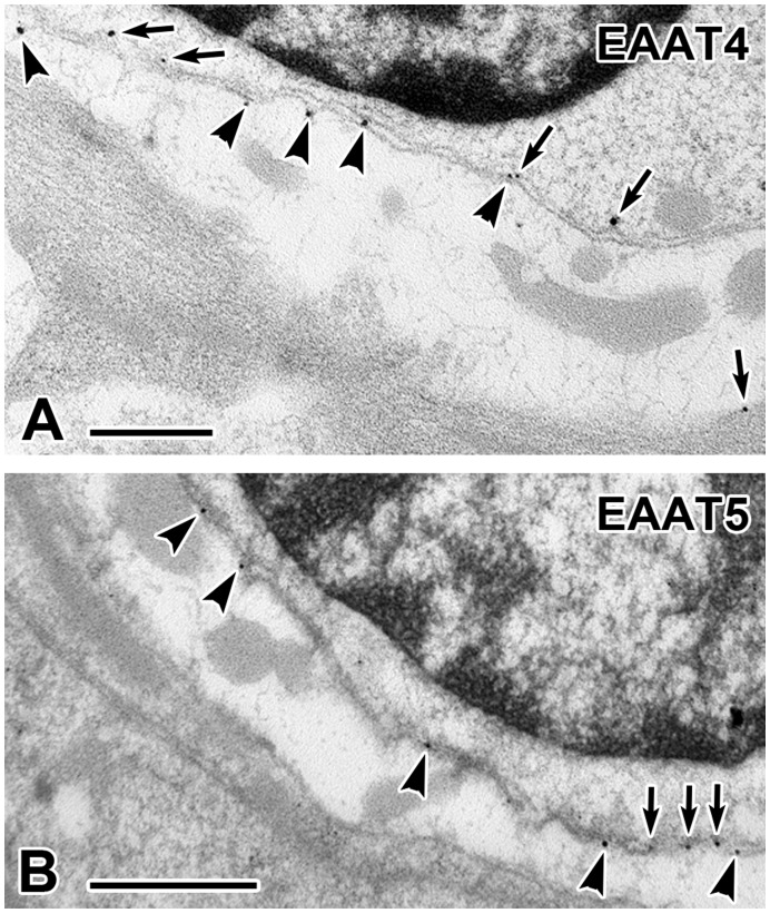
Figure 5. EAAT4 and EAAT5 protein localization in mouse tissue.
Electron micrographs of EAAT4 (A) and EAAT5 (B) immunogold labeling particles on hair-cell membrane (arrows), calyx inner-face membrane (arrowheads) and calyx outer-face membrane (arrow, lower right in A). In both panels from top to bottom, the darkened area is a hair-cell nucleus rimmed by hair-cell cytoplasm, hair-cell and calyx inner-face membranes. The lightened area with gray mitochondria is a calyx ending whose outer-face abuts supporting cells. Scale bars: 0.5 µm.

