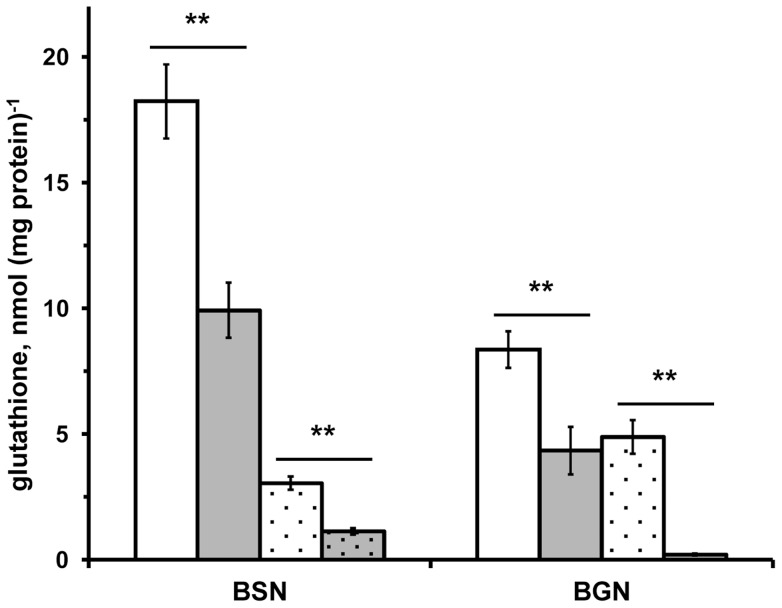
Figure 2. Glutathione in A. vinelandii.
(A) Glutathione was detected in wild-type (UW136; white bars) and in RhdA-null mutant (MV474; grey bars) A. vinelandii strains grown in Burk's medium containing sucrose (BSN) or gluconate (BGN) as carbon source in the absence (not-dotted bars) and in the presence (dotted bars) of phenazine methosulfate (PMS). Measured glutathione includes both the total reduced-glutathione and the DTT-reducible fraction of total free glutathione and is expressed as function of the protein amount detected in 10 mM Tris-HCl 0.1 M NaCl (pH 8) extracts of cell-samples from the same growths. All data are the mean of three independent replicates ± standard deviation. Differences significant for P<0.01 (**) are indicated (Student's t test).

