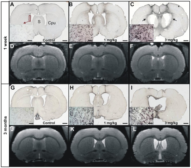
Figure 4. Evaluation of the astrogliosis in control (A, G), 1 mg/kg (B, H) and 3 mg/kg (C, I) icv-streptozotocin treated groups, seven days (A–C) and three months (G–I) post injection.
MRI sections corresponding to the histological sections are shown in D–F (one week post- injection) and J–L (three months post-injection). Low and high (left insets) magnification images showing Glial Fibrillary Acidic Protein (GFAP) staining. A severe astrogliosis was detected in the 3 mg/kg treated group (halo of activated astrocytes in C (arrow), insets in C and I). A slight astrogliosis could be detected in the 1 mg/kg animals (B, H). The inflammation process was not obvious on MR images at both times and in all groups (D–F for one week post-injection, and J–L for three months post-injection). However, dilation of the ventricles was clearly visible on these same images in STZ-treated-animals. S: Septum. Cpu: Striatum. Scale bars for low magnification images = 2 mm and scale bars for insets = 50 µm.

