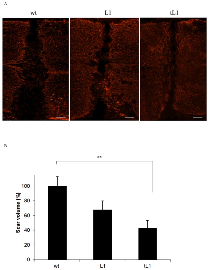
Figure 3. Transplantation of tL1 NSCs reduces lesion volume after spinal cord injury.
The lesion sites are delineated by glial fibrillary acidic protein (GFAP) expressing astrocytes (red) six weeks after transplantation of tL1, L1 or wt NSCs. Five mice per group were analyzed. Shown are mean values with SEM from Image J. The scar volume in the wt NSC group was set to 100%. Statistical analysis was performed by one-way ANOVA with Tukey's post-hoc test (** p<0.01). Scale bar = 150 µm.

