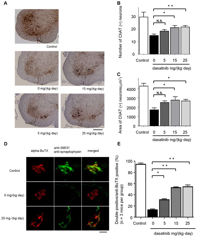
Figure 6. The effect of dasatinib on motor neuron survival in G93A mice.
A: Spinal cord (L1-3) specimens from 120-day-old mice were immunostained with anti-ChAT antibody. The mice were administered the indicated amounts of dasatinib daily from postnatal day 56 to day 120 (n = 8 mice per group). Scale bar: 250 µm. B: The number of ChAT-positive neurons in the sections described in Fig. 6A was counted using Image J software. Dasatinib prevented the loss of ChAT-positive motor neurons in the ventral horn of G93A mice at doses of 15 mg/(kg·day) (P<0.05) and 25 mg/(kg·day) (P<0.01). Statistics were evaluated using 1-way ANOVA with Dunnett's post-hoc test. *P<0.05, **P<0.01. C: The area of ChAT-positive neurons in the sections described in Fig. 6A was determined using Image J software. Dasatinib increased the size of motor neuron cell bodies at doses of 15 and 25 mg/(kg·day) (P<0.05). Statistics were evaluated using 1-way ANOVA with Dunnett's post-hoc test. *P<0.05. D: To investigate the innervation status of NMJs, frozen quadriceps femoris specimens from 120-day-old mice were stained with alpha-BuTX (red) and anti-synaptophysin (green) or anti-SMI31 (green) antibodies. Representative NMJs visualized with the confocal laser scanning microscopy are shown. The mice were administered the indicated amounts of dasatinib daily from postnatal day 56 to day 120. Scale bar: 10 µm. E: The ratio of double-immunostained innervated NMJs to total NMJs is summarized. One hundred immunostained NMJs were investigated in each dasatinib-treated mouse (n = 3 mice per group). Dasatinib significantly ameliorated the destruction of NMJ innervation in G93A mice at doses of 5 (P<0.05), 15, and 25 mg/(kg·day) (P<0.01). Statistics were evaluated using 1-way ANOVA with Dunnett's post-hoc test. *P<0.05, **P<0.01.

