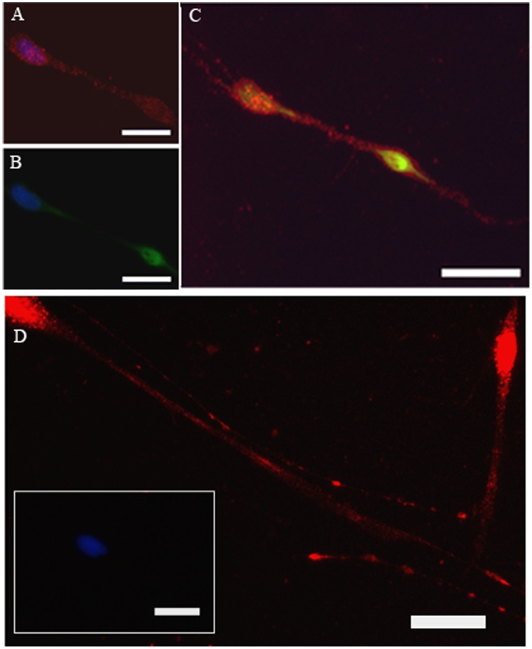
Figure 1. β2 receptors are identified by anti-β2 receptors antibody on human trachea parasympathetic neurons (red, A, B–D) under high (A,C) and low (D) power.
Neurons are labeled with anti-neurofilament antibodis (B, green) and the merged image (for neuronal and β2 receptor staining) is shown in C. Nuclei stain blue with DAPI. The insert of D is the absence of primary antibody. Magnification bars: 50 µm.

